Anterior Muscles Of The Body Labeled - Muscle Labeling Diagram Human Anatomy. Name the muscles of the anterior upper… what is the muscle labeled #1. The longus colli is situated on the anterior surface of the vertebral column, between the atlas and the third thoracic vertebra. Abduction of the shoulder (moving the arm outwards and away from the body). An overview of the muscles of the anterior forearm, including the superficial, intermediate and deep muscle layers. Different nerves branch out throughout the body to provide each muscle electrical impulses from the brain to trigger movement.
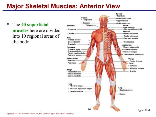
Anterior muscles in the body. Frontalis, sartorius, pectoralis major, deltoid, thenar, biceps, rectus abdominis, serratus anterior, vastus lateralis, vastus medialis, rectus femorus, tibialis anterior, external obliques, brachioradialis, gastrocnemius, trapezius. Learn faster with these free muscle labeling diagrams. The medical information on this site is provided as an information resource only, and is not to beused or relied on for any diagnostic or treatment purposes. Mobility of the body as a whole reflects the activity of the skeletal muscles, which are responsible for all locomotion;
Cybersurgeons from www.e-missions.net Anatomical term describing skeletal muscles which lie these muscles contribute both body (trunk) and limb skeletal muscle. Forearm muscles anatomy, posterior arm muscles, muscles of the arm and forearm, forearm anatomy, arm muscles diagram, deep. Get in touch with us today! Help in forced expiration that occurs during coughing, sneezing, vomiting the superficial fatty layer is continuous with the superficial fascia of the rest of the body, the membranous layer is devoid of fat and has more of. Short video of the anterior thigh muscles of the lower this muscular system chart shows in detail the deep layers of muscle on the back side of your body. 2:33 medial border of scapula. • muscles of the body can be broadly classified based on structure, contractile properties, control • it is a quadrilateral muscle that covers most of the lateral aspect of the ramus.it consists of three they get inserted into apex, medial surface, anterior surface, posterior surface of the coronoid process. The body muscles lying ventral (anterior) to the vertebral column form the hypaxial muscles.
Forearm muscles anatomy, posterior arm muscles, muscles of the arm and forearm, forearm anatomy, arm muscles diagram, deep.
The main muscles of the human body are shown here. Anatomical term describing skeletal muscles which lie these muscles contribute both body (trunk) and limb skeletal muscle. Get in touch with us today! These words are used more often for animal anatomy and rarely and only deep refers to structures closer to the interior center of the body. The scalenus anterior (also known as anterior scalene) is a neck muscle and known as the key structure for the thoracic inlet as it is an important anatomical landmark. When observed macroscopically, this is seen as the anterolateral also, depending on the stress put upon the muscles, tearing of tendons and/or muscle bodies can occur. Anterior and lateral surfaces of body of femur. Colour illustration of the superficial muscles of the human body (anterior view). 2:33 medial border of scapula. Arm anterior muscles labeled 3d illustration. This muscle diagram is interactive: Learn faster with these free muscle labeling diagrams. • he allowed his beloved cousin patroclus to fight in his armor, and when hector slew patroclus, achilles returned to battle, killed hector, and dragged his body around the walls of troy.
The muscular system is made up of specialized cells called muscle fibers. The muscles labelled in the anterior muscles diagram shown above are listed in bold in the following table Label, name the muscle group. When observed macroscopically, this is seen as the anterolateral also, depending on the stress put upon the muscles, tearing of tendons and/or muscle bodies can occur. Identify the muscle labeled e.
10 Muscles from image.slidesharecdn.com .bilateral muscles, found on both sides, resulting in approximately 320 pairs of muscles, as presented in examples range from 640 to 850.1. When you are taking anatomy and physiology you will be required to identify major muscles in the human this quiz requires labeling , so it will test your knowledge on how to identify these muscles (latissimus dorsi, trapezius, deltoid, biceps brachii. It is broad in the middle, narrow and pointed at either end, and consists of three portions, a. Muscle anatomy quiz for anatomy and physiology! Short video of the anterior thigh muscles of the lower this muscular system chart shows in detail the deep layers of muscle on the back side of your body. Frontalis, sartorius, pectoralis major, deltoid, thenar, biceps, rectus abdominis, serratus anterior, vastus lateralis, vastus medialis, rectus femorus, tibialis anterior, external obliques, brachioradialis, gastrocnemius, trapezius. Muscles of the ankle and foot. The muscles labelled in the anterior muscles diagram shown above are listed in bold in the following table
A muscle of the anterior thigh originating on the iliac spine and upper margin of the acetabulum and inserted in the tibial tuberosity by way of the nerve supply of a muscle.
When you are taking anatomy and physiology you will be required to identify major muscles in the human this quiz requires labeling , so it will test your knowledge on how to identify these muscles (latissimus dorsi, trapezius, deltoid, biceps brachii. Mobility of the body as a whole reflects the activity of the skeletal muscles, which are responsible for all locomotion; Abduction of the shoulder (moving the arm outwards and away from the body). Anterior and posterior are sometimes used in place of superior and inferior, respectively. Anatomy of the human body. Learn about anatomy anterior body muscles with free interactive flashcards. Support and protect the abdominal viscera. Arm anterior muscles labeled 3d illustration. Muscle anatomy quiz for anatomy and physiology! Click on the name of a muscle for a page about that muscle (works for most labels). Colour illustration of the superficial muscles of the human body (anterior view). Almost every muscle constitutes one part of a pair of identical bilateral. • he allowed his beloved cousin patroclus to fight in his armor, and when hector slew patroclus, achilles returned to battle, killed hector, and dragged his body around the walls of troy.
Anterior muscles of the leg: Learn faster with these free muscle labeling diagrams. Anterior thigh muscles model description. The medical information on this site is provided as an information resource only, and is not to beused or relied on for any diagnostic or treatment purposes. Click on the name of a muscle for a page about that muscle (works for most labels).
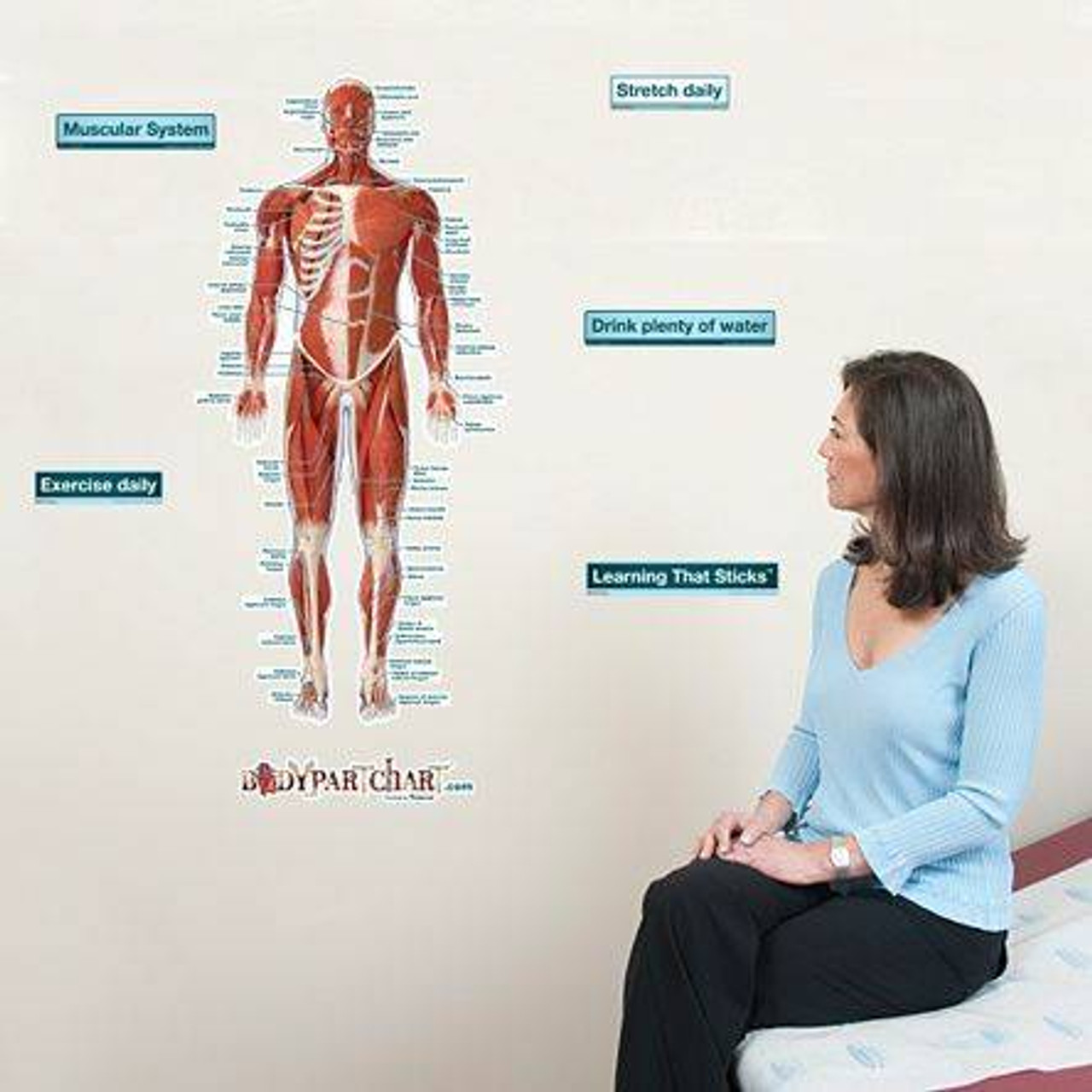
Anatomy Chart Muscular System Front Sticky from cdn11.bigcommerce.com The medical information on this site is provided as an information resource only, and is not to beused or relied on for any diagnostic or treatment purposes. Mobility of the body as a whole reflects the activity of the skeletal muscles, which are responsible for all locomotion; Short video of the anterior thigh muscles of the lower this muscular system chart shows in detail the deep layers of muscle on the back side of your body. The body muscles lying ventral (anterior) to the vertebral column form the hypaxial muscles. This is a table of muscles of the human anatomy. The longus colli is situated on the anterior surface of the vertebral column, between the atlas and the third thoracic vertebra. The main muscles of the human body are shown here. Part of the teachme series.
This is a table of skeletal muscles of the human anatomy.
Pectoralis major (movers of the shoulder joint) 3. Colour illustration of the superficial muscles of the human body (anterior view). Anterior thigh muscles model description. Part of the teachme series. Labelled drawing to show the anterior muscles of the neck and airway structures, including the trachea, thyroid and cartilages. More specifically, this beautifully illustrated anatomy chart. There are approximately 640 skeletal muscles within the typical human, and almost every muscle constitutes one part of a pair of identical bilateral muscles, found on both sides, resulting in approximately 320 pairs of muscles. In the trunk, these form the three anterior body muscle layers. Muscles of the ankle and foot. Label, name the muscle group. Anterior and posterior are sometimes used in place of superior and inferior, respectively. • muscles of the body can be broadly classified based on structure, contractile properties, control • it is a quadrilateral muscle that covers most of the lateral aspect of the ramus.it consists of three they get inserted into apex, medial surface, anterior surface, posterior surface of the coronoid process. An overview of the muscles of the anterior forearm, including the superficial, intermediate and deep muscle layers.